Resource library
Practical teaching assets for interventional pulmonology educators, learners, and clinician-builders.
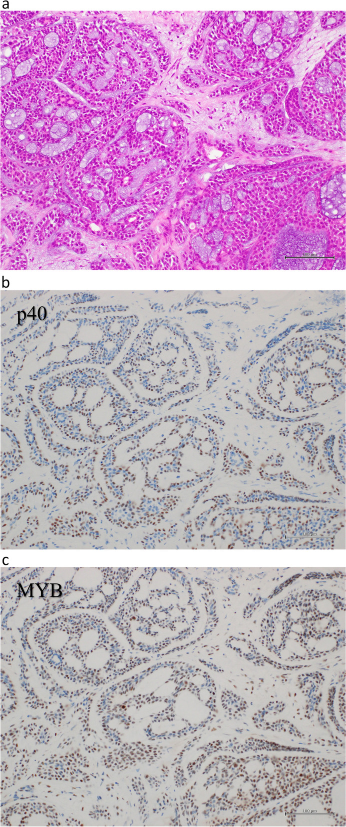
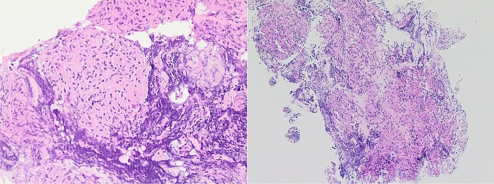
Adenoid cystic carcinoma (cribriform pattern, p40 and MYB IHC)

Adenoid Cystic Carcinoma (Cribriform Subtype)

Cribriform tumor structure (H&E 40× and 200×)

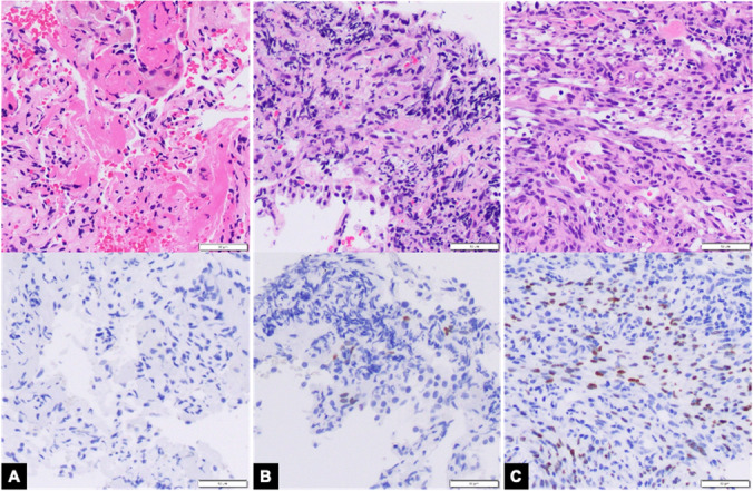
Cytokeratin immunohistochemistry (negative)

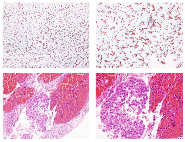
EBUS thyroid aspiration (poorly differentiated adenocarcinoma, TTF1/CEA/CK7 IHC)

EBUS-FNA squamous cell carcinoma (H&E, p40 and CK14 IHC)

Endobronchial lipoma (mature adipose tissue with mucoid changes, H&E)

Endobronchial tumor (polypoid lesion, H&E with myxoid stroma)
Endobronchial tumor (pre- and post-ablation biopsy comparison, H&E)
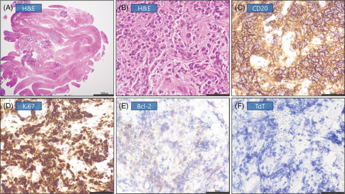
Immunohistochemistry panel (CD20, Ki67, Bcl-2, TdT)
Kaposi Sarcoma (Forceps vs Cryobiopsy vs FNA)

Lung cryobiopsy (H&E and ex-vivo confocal microscopy)

Lymph node squamous cell carcinoma (H&E low magnification)

Malignant mesothelioma (epithelioid and sarcomatoid, WT1 and calretinin IHC)
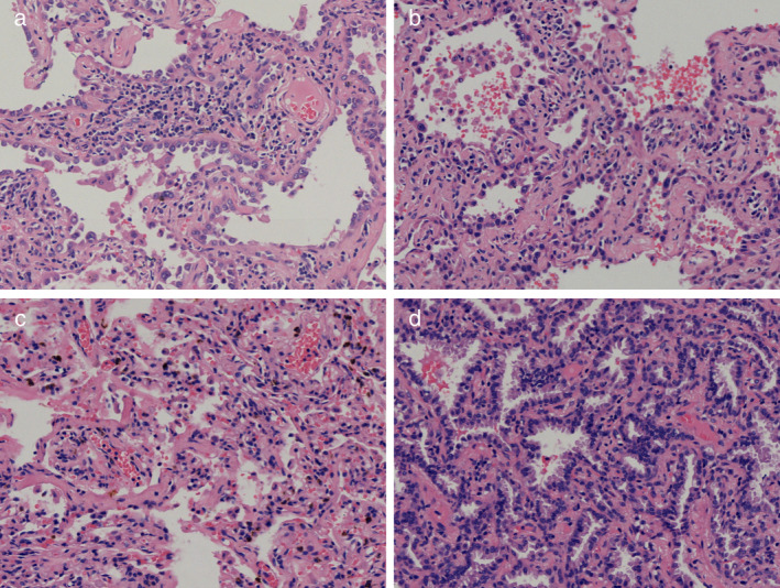
Multiple lung nodules (AIS, MIA, chronic inflammation, H&E)

PLCH vs HP (fibromyxoid foci and interstitial fibrosis)

Pleural Endometriosis with Stromal Invasion

Pleuropulmonary blastoma (chondrosarcoma, glandular and blastemal components, H&E)

S-100 immunohistochemistry (positive)
Sarcoidosis granuloma (cytoblock with Crown Cut needle)

Tissue core and blood contamination evaluation (H&E slides)


Tracheal schwannoma (H&E with Antoni A/B areas, nuclear palisading)

Tracheal Schwannoma (S100 Positive)

Transbronchial biopsy adenocarcinoma (RUL with ROSE, H&E)
Cytology – Adenocarcinoma cells (high N/C ratio, TTF1 IHC)
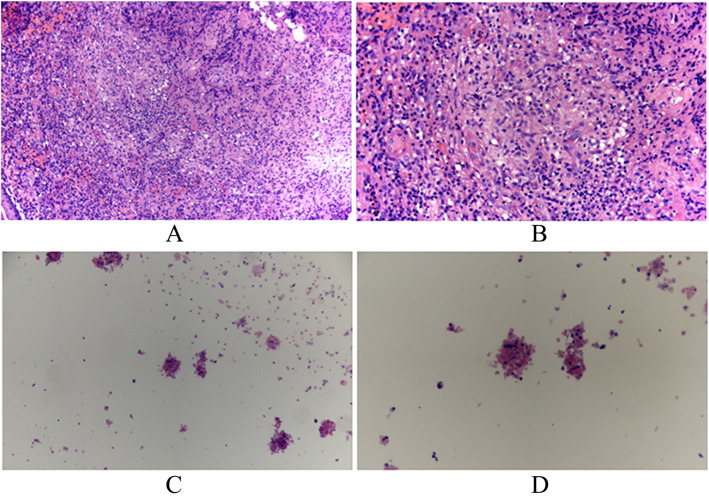
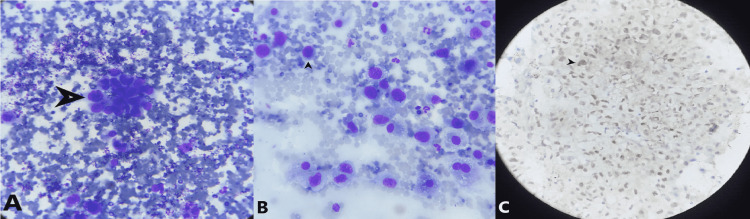
Cytology – Aspirated pus (epithelioid cells, lymphocytes, granuloma formation)
Cytology – EBUS-FNA cell block vs smear (subcarinal metastatic adenocarcinoma)

Cytology – Lymphocytes in aspirated material (H&E)

Cytology – ROSE classification (Class 3 vs Class 4 cluster density)

Cytology – ROSE from EBUS-FNA (22G Mediglobe needle)

ROSE/Pathology Comparison – Forceps vs cryobiopsy (metastatic urothelial carcinoma and adenocarcinoma)

Specimen – Resected lung mass (RLL and RML)
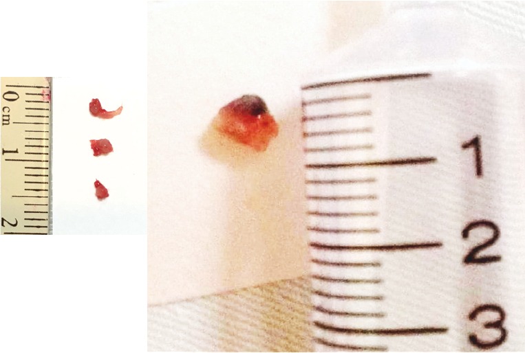
Specimen Comparison – Forceps biopsy vs cryobiopsy
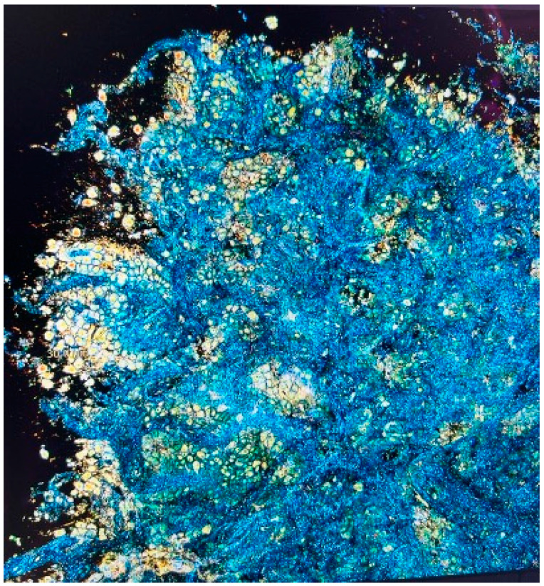
FFOCT-DCI – Technique description (high-resolution 3D biopsy imaging)

Digital Pathology – Quantitative analysis workflow (calibration and threshold measurement)

CT/Cytology – Mediastinal lymphadenopathy (small cell lung carcinoma, H&E)

CT/Cytology – Mediastinal mass with necrosis (EBUS-TBNA adenocarcinoma)

CT/Pathology – Discordant histology (LUL mass, SCC vs adenosquamous carcinoma)

Confocal Laser Endomicroscopy – Squamous cell carcinoma vs adenocarcinoma (with cytology)