Resource library
Practical teaching assets for interventional pulmonology educators, learners, and clinician-builders.

Pleural Cryobiopsy vs Forceps (Tissue Integrity/Size)

Endometriosis (Visceral and Parietal; Lesion Types)
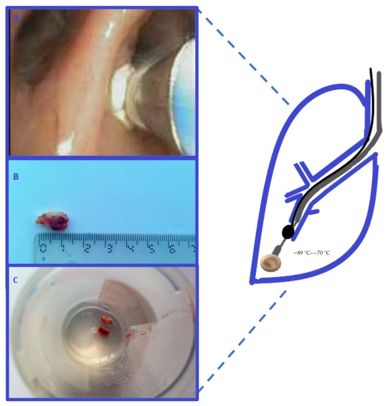
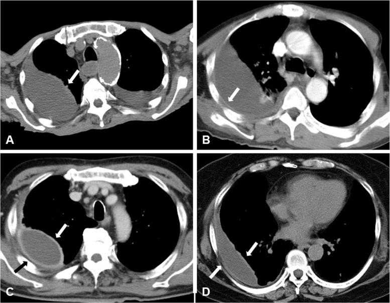
Medical Thoracoscopy – Cryobiopsy (Peripheral Tumor)

Medical Thoracoscopy – Precut Biopsy (Stepwise)
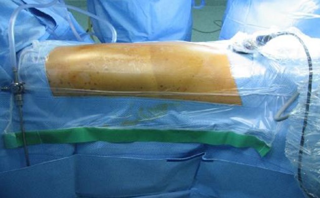
Patient Positioning (Thoracoscopy)

Precut Step‑Up Biopsy Strategy (Schematic)

Thoracoscopic Appearances (Solid/Fibrous/Thickened)

Thoracoscopy + Urokinase Fibrinolysis (Intrapleural Nets)
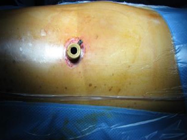
Trocar Placement (7th Intercostal Space)
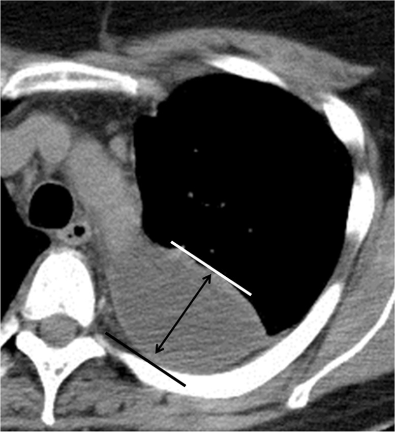

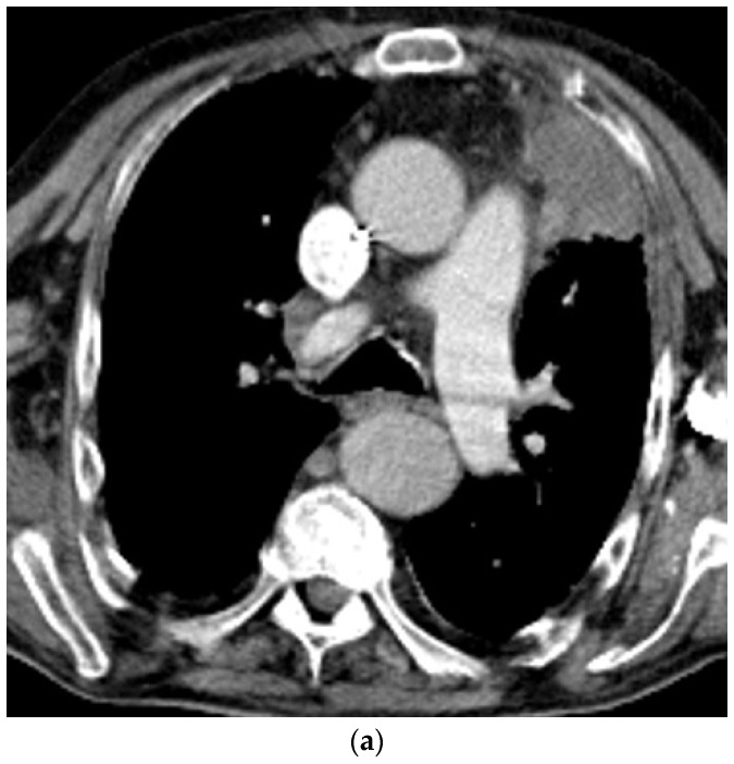
Pleura/Vascular Tumor – CT/MR & pathology

Pleuroscopy – Biphasic mesothelioma and pleural metastasis (LTF-Y0032 with 180° curvature)

Pleuroscopy – Empyema (thick loculated with fibrin membrane)

Pleuroscopy – Lesion distribution map (visceral and parietal locations by segments)

Pleuroscopy – Malignant mesothelioma and lymphoma (NBI with vascular patterns)

Pleuroscopy Equipment – Conventional vs new pleuroscope comparison (biopsy forceps positioning)
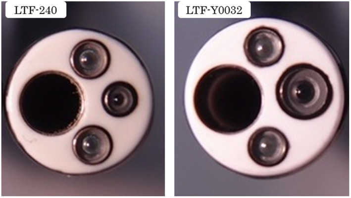
Pleuroscopy Equipment – Diameter comparison (LTF-240 vs LTF-Y0032)

Pleuroscopy/Bronchoscopy – Pleural adhesions and whitish mass (right bronchus intermedius)

Pleuroscopy/Pathology – Normal parietal pleura pCLE features (chia seed sign, H&E correlation)

Pleuroscopy/Pathology – pCLE features (pleural metastasis with H&E correlation)
CT/Ultrasound/CEUS – Epithelioid mesothelioma (bronchial arterial enhancement, washout)
CT/Ultrasound/CEUS – Epithelioid mesothelioma (circular pleural thickening, systemic arterial enhancement)
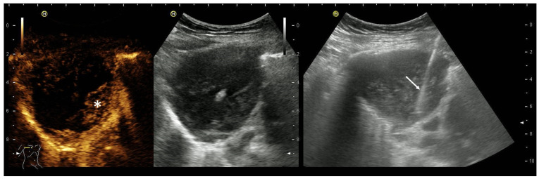
CEUS – Subpleural lung tumor biopsy (needle positioning in vital tissue)

Chest X-ray/CT/Pleuroscopy – Precut and cryobiopsy technique (adenocarcinoma with HE staining)

Chest X-ray/CT/Pleuroscopy – Precut technique (biphasic mesothelioma biopsy with CAM/HE staining)

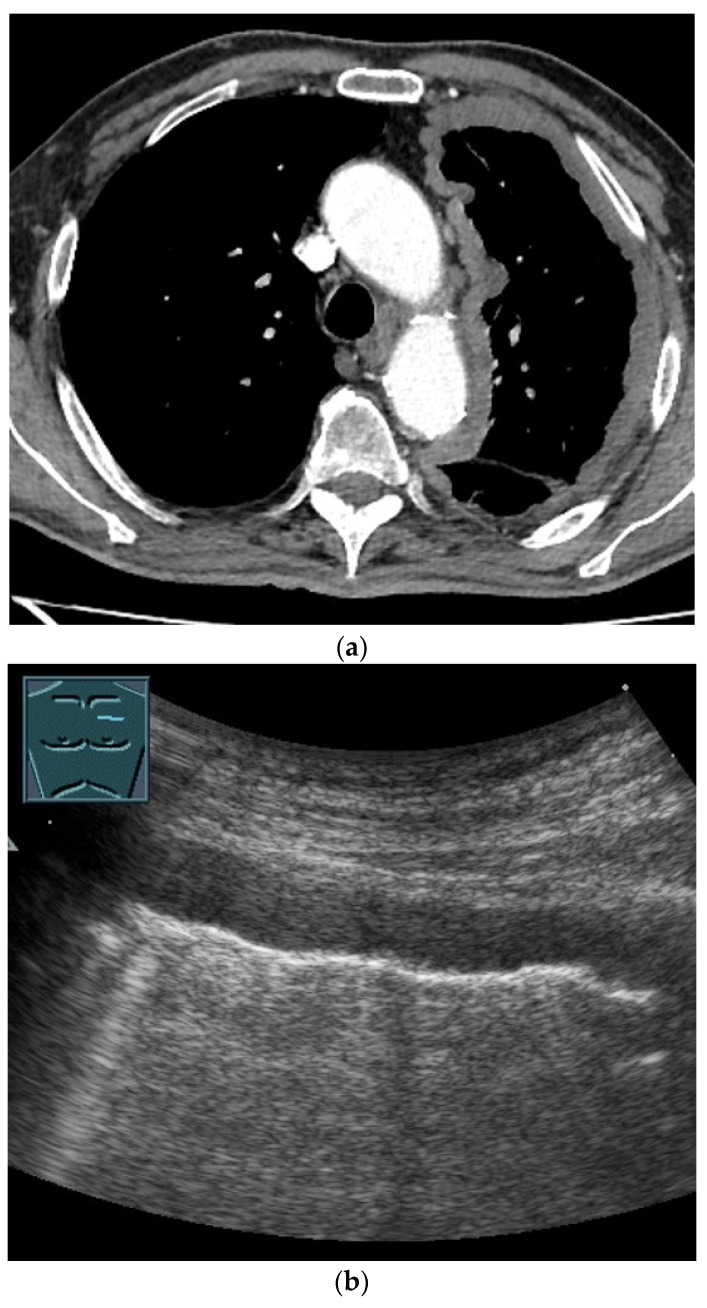
Ultrasound/Pleuroscopy – Pleural effusion patterns (anechoic, complex, septated, homogeneous)

Ultrasound/Pleuroscopy Correlation – Pleural sliding sign, effusion, and adhesions

Ultrasound – Pleural biopsy technique (target identification, vascularization assessment)

CT/Pleuroscopy Correlation – Pleural nodules (lung cancer metastasis vs tuberculous pleurisy)