Resource library
Practical teaching assets for interventional pulmonology educators, learners, and clinician-builders.

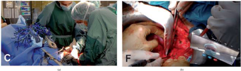
RATS – Azygos vein exposure


RATS – Post‑azygos division

Conventional VAL-MAP technique (step-by-step)

CT-guided cyanoacrylate localization

CT-guided indocyanine green localization

CT-guided memory alloy coil localization

Lungpro-assisted metallic marker localization

RFID marker placement (wedge resection steps)

VAL-MAP 2.0 technique (step-by-step)
Intraoperative Imaging – ICG dye localization (VATS with fiducial marker)

Intraoperative Imaging – NIR fluorescence with ICG (Firefly mode vs white-light)

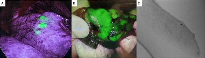
Fluoroscopy/Pathology – Lung nodule resection confirmation (ICG-coil marker, specimen)

Forensics – Tracheal ring injuries (autopsy)

Magnetic Navigation – Pulmonary nodule localization (RUL, methylene blue, thoracoscopic verification)
Telemedicine – Intra‑op 3D anatomy & consult

Ultrasound – Intraoperative pulmonary nodule localization

VAL-MAP – Dual dye technique (indigo carmine and ICG with NIR thoracoscopy)
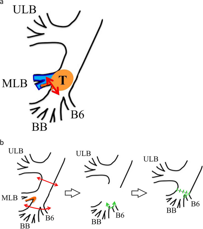
Diagram – Endobronchial tumor resection (electrocautery snare and RML sleeve lobectomy)
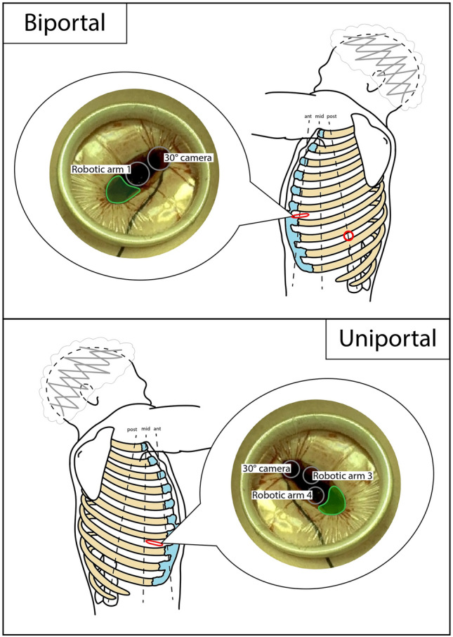
Diagram – RATS biportal and uniportal access positioning (schematic)
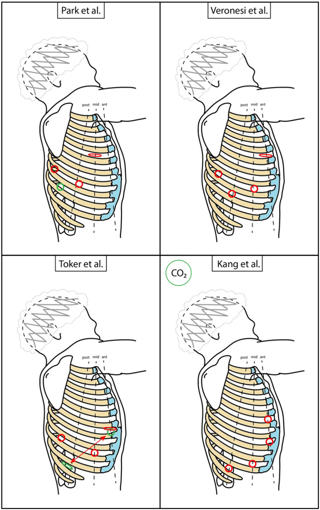
Diagram – RATS main access positioning (schematic with mini-thoracotomy)
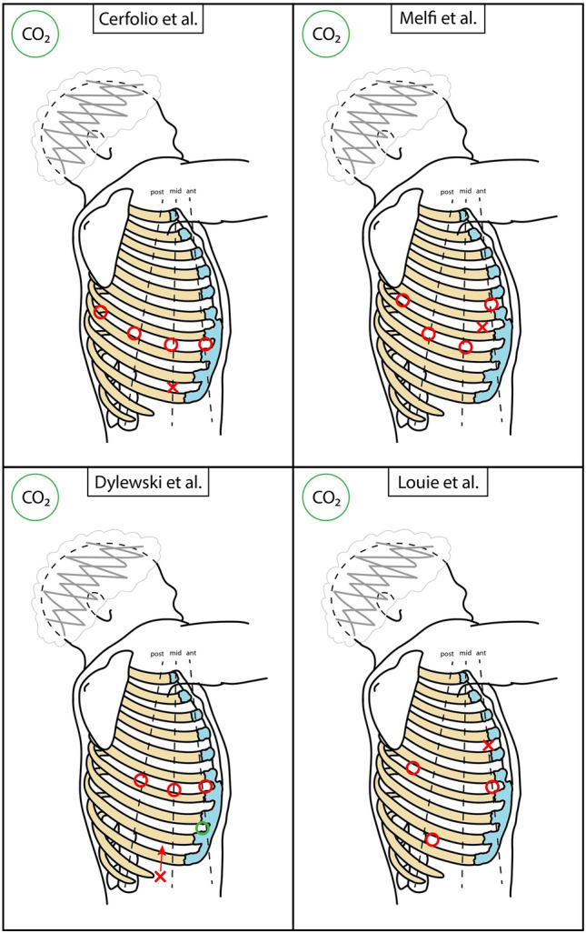
Diagram – RATS main access positioning (schematic with tunneling)
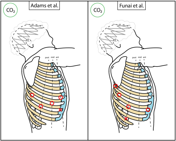
Diagram – RATS main access positioning (schematic)

Diagram – VAL-MAP 2.0 workflow (microcoil placement, 3D reconstruction, fluoroscopy-guided resection)


CT/PET – Pulmonary lesion (surgical port mapping)