Resource library
Practical teaching assets for interventional pulmonology educators, learners, and clinician-builders.
CT/Bronchoscopy – Tracheal esophageal tumor (stent obstruction, APC recanalization)

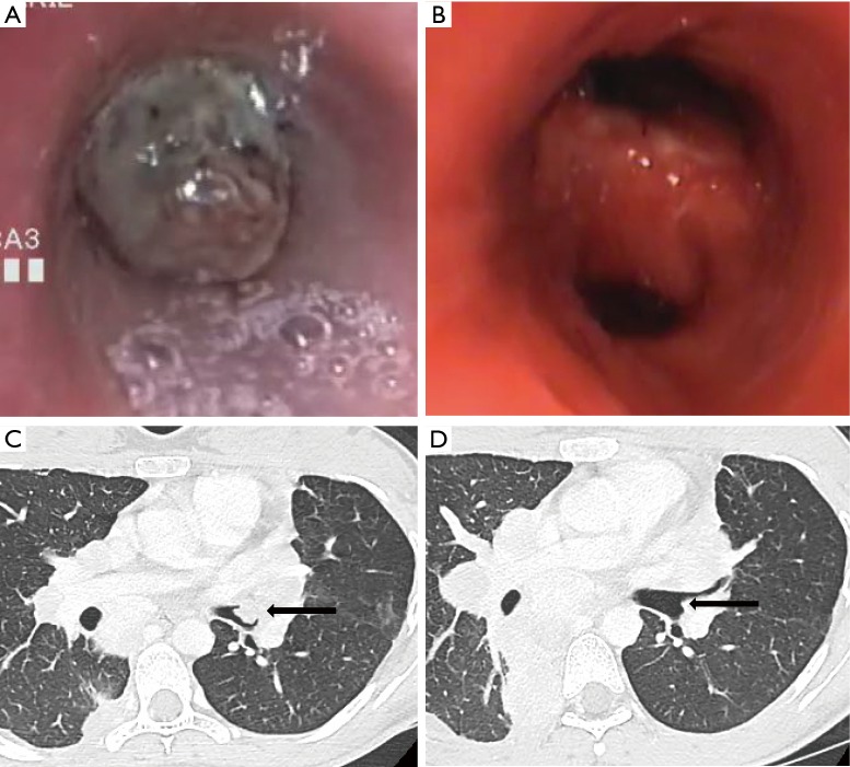
CT/Bronchoscopy – Tracheal polypoid metastasis (rigid bronchoscopy coring, APC)

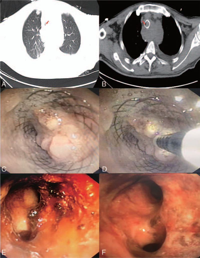
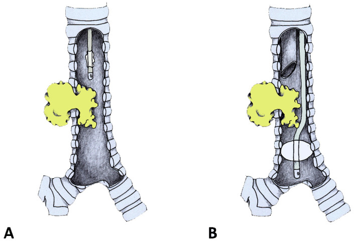
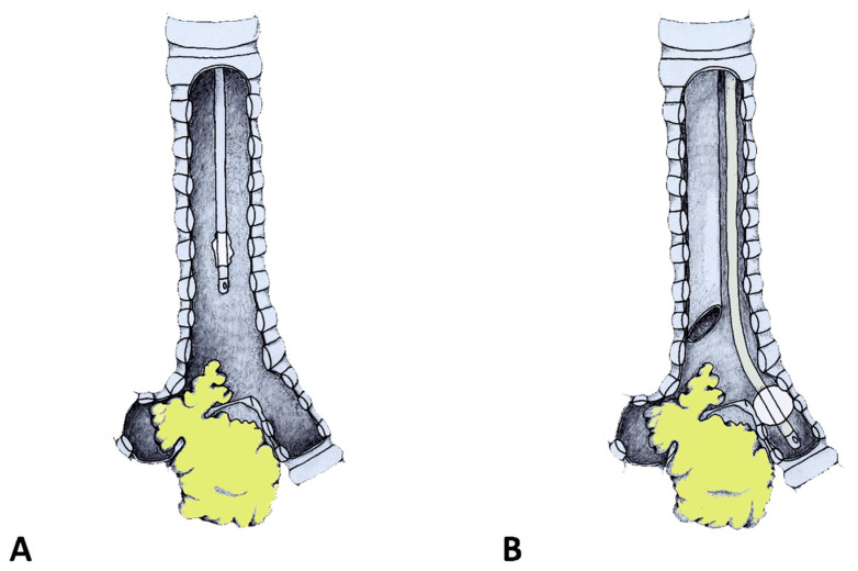
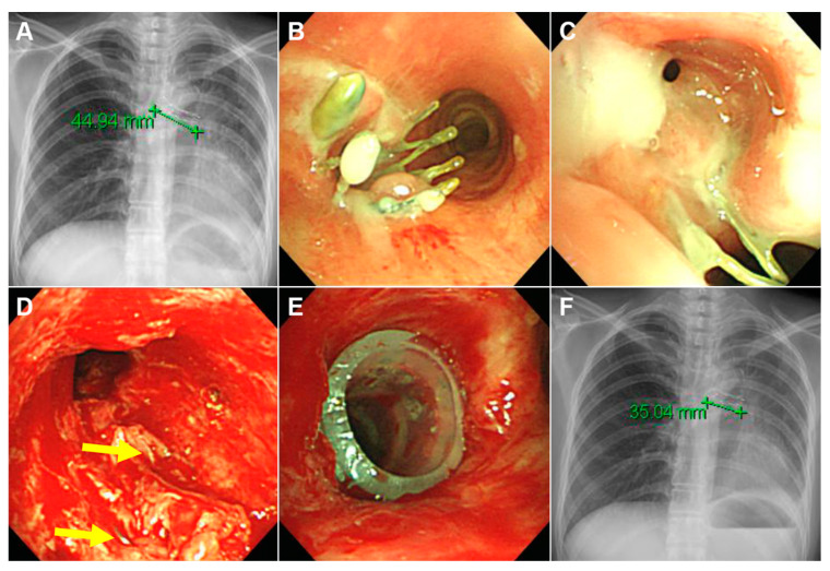
CT/Bronchoscopy – Tracheoesophageal fistula (SEMS placement and removal, stenosis management)

CT/Bronchoscopy Correlation – Tracheal mass with near-complete obstruction

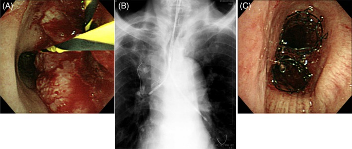
CT/Bronchoscopy Correlation – Tracheoesophageal fistula (esophageal tumor, stent placement with barium swallow)

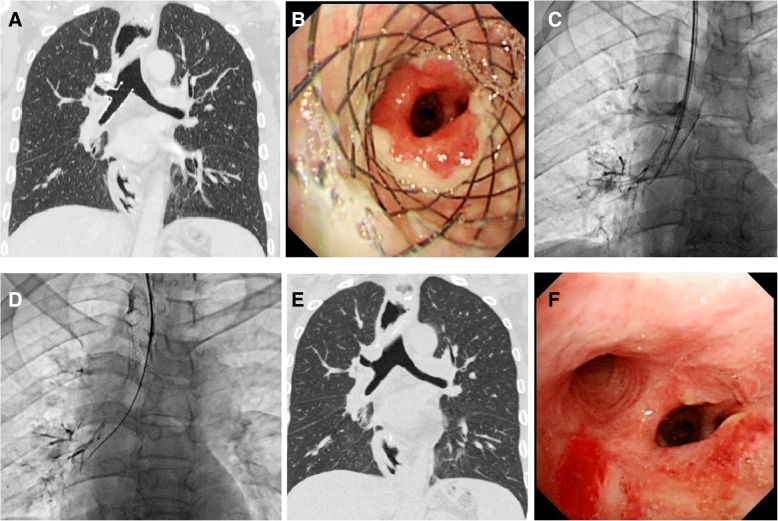
CT/Bronchoscopy/Chest X-ray – LMB compression (thoracic aortic aneurysm, stent placement timeline)

CT/Bronchoscopy/Chest X-ray – Mediastinal mass (LMB occlusion, stent placement)

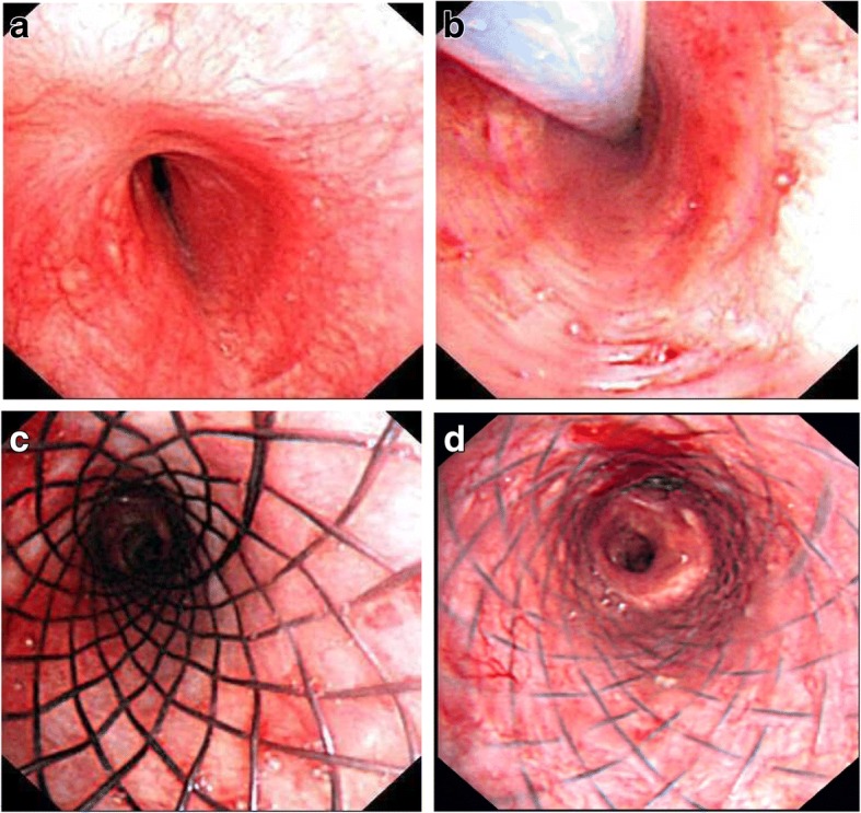
Silicon stent follow-up (6-month, removal with persistent stenosis)

Single‑Use Bronchoscope (Airway Burn/Cryotherapy)

Subglottic stenosis (balloon dilation, Kenalog injection, follow-up)

Subglottic stenosis (IT knife palliation)

Subglottic stenosis (needle knife incisions, Kenalog injection)

Tracheal adenoid cystic carcinoma (pre/post resection, follow-up)

Tracheal and LMB obstruction (stent placement, proximal and distal views)

Tracheal compression (rigid bronchoscopy dilation, Dumon Y stent at carina)

Tracheal invasion and perforation (silicon Y-stent placement for fistula)

Tracheal lobulated mass (pre/post removal, 12-month follow-up)

Tracheal MAO (malignant melanoma, Polyflex stent)

Tracheal papilloma (pre/post endobronchial treatment)

Tracheal polypoid tumor (APC, cryoablation, snare resection sequence)

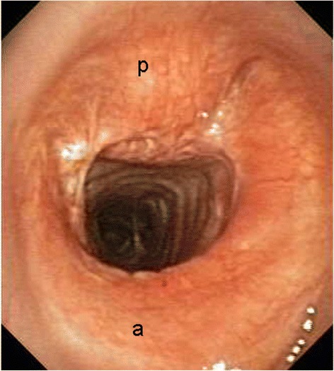
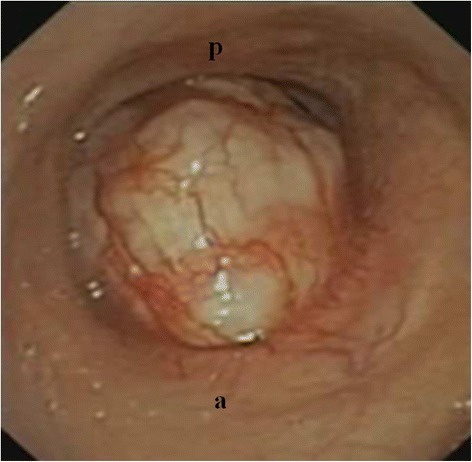
Tracheal Schwannoma (White Pedunculated Lesion)

Tracheal squamous cell carcinoma (diode laser coagulation, scabbard trachea)

Tracheal stenosis (covered Ultraflex for malignant lymphoma)


Tracheoesophageal fistula (covered Ultraflex with migration)

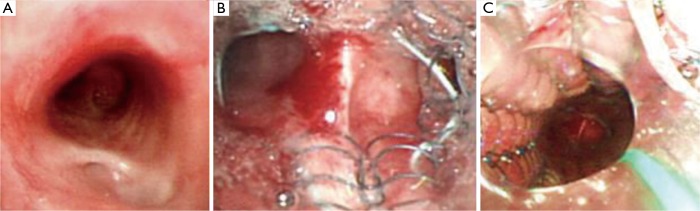
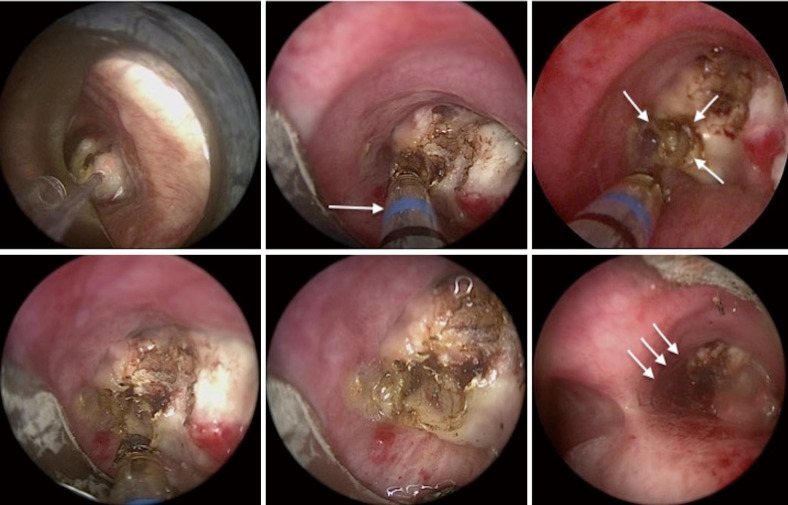
Tracheoesophageal fistula suturing (rigid bronchoscopy technique)

Tumor clearance (mechanical removal via rigid bronchoscope)

Types of Malignant Airway Obstruction (Examples)
Typical Carcinoid (Pre/Post Laser Resection)

Y-shaped stent removal (failed expansion, hook retrieval)
Bronchoscopic Images/CT – Dumon Oki stent (RMB extrinsic compression, post-stenting)
Bronchoscopic Images/CT – RML fistula stent management (placement, restenosis, removal timeline)

Bronchoscopic Images/CT – Tracheal stenosis (covered Ultraflex for lymphoma, R-CHOP therapy, 6-year survival)

Bronchoscopic Images/CT – Tracheal stenosis (spiral Z stent for thymic cancer, 6-year survival)

Cryo-debulking (endoluminal tumor)

Cryotherapy (silicone stent-induced granulation)


Dynamic stenosis and stent complications (Y-silicone stent, metallic stent, granulation)

Early Endobronchial Lesion (Silver Hue)

Early squamous cell carcinoma (central airways)

Endobronchial hamartoma (core-out and cryotherapy sequence)

Endobronchial leiomyoma (LMB obstruction, EC snare/cryo debulking)

Endobronchial lipoma (cryorecanalization sequence)
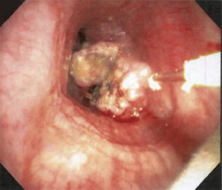
Endobronchial Mass (Tracheal Lesion at Presentation)

Endobronchial mucous adenoma (treatment timeline with histology)
Endobronchial tumor (middle lobe, pre/post resection)

Endoluminal Carcinoid (Resection Correlation)

Extrinsic CAO (silicone Y-stent for airway recanalization)


Fibroepithelial tumor (LUL superior segment, pre/post treatment)

Fibrotic Stenosis (Left Main Bronchus)

Fogarty Balloon (Hemoptysis Control)


Foreign body removal (intermediate bronchus, granulation cautery, basket/forceps techniques)
FOT placement above stenosis (rigid scope insertion with cuff)
FOT placement for carinal obstruction (cuff inflation technique)


iSGS proximal stenosis progression (cross-sectional evolution stages)


LMB occlusion management (microdebrider debulking, APC, stent placement)
LMB stenosis (nitinol stent, 7-year follow-up)

MAO bilateral main bronchi (pre/post therapy, Y-stent)

Mediastinitis with Tracheal Fistula Drainage

Microwave Ablation (LMS Tumor)

Mixed CAO (cryo-debulking and stent placement)

Mixed CAO Resected (Airway Restored)

Modified silicone Y stent removal (post-tuberculous destroyed lung)

Modified Y stent placement (bronchiectasis, occluded LUL branch)

Montgomery T-tube placement (subglottic stenosis, pre/post)

Multimodal CAO management (carinal SCC, laser, Y stent, radiation)

Multimodal MCAO management (bilateral stent placement with spray cryotherapy)

Neoplasia in airway stent (endoscopic resection sequence)

Paratracheal tumor excision (covered stent with balloon dilation)

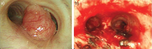
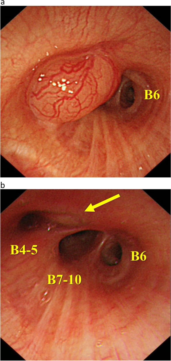
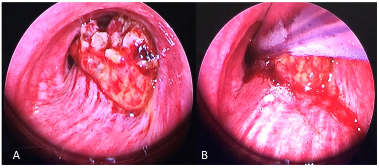
Polypoid lesion (RMB vascularized tumor)

Postoperative timeline (pre-op to 3.5 years)

Post‑Transplant Airway Complication Types

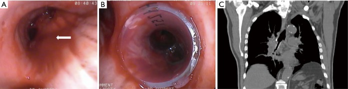
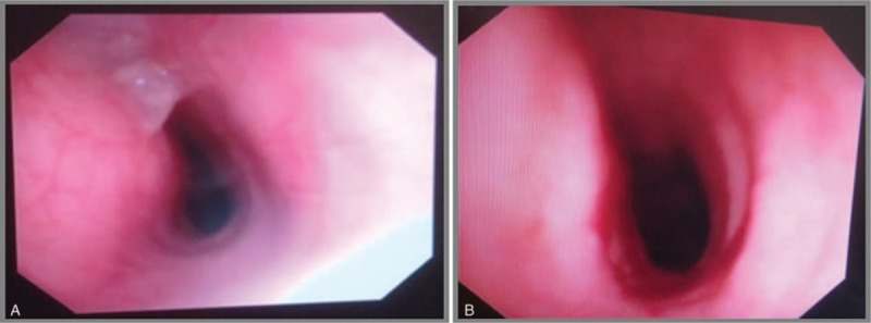
Recanalization Post‑Procedure (Trachea)

Rigid bronchoscope and suction catheter (carina level)
SEMS for tracheoesophageal fistula (mechanical ventilation, no fluoroscopy)

SEMS removal (embedded stent, laser vaporization, Y-shape SEMS, PTTS)

Silicon stent follow-up (1-month, granulation formation)

Silicon stent follow-up (4-month, increased granulation)

3D printed stent (customized silicone for complex stenosis)
Airway metastasis (LMB, hepatocellular carcinoma, laser/mechanical debulking)

APC and diode laser (endobronchial treatment sequence)

Balloon Dilation (Incisions → Rigid Scope)

Balloon Dilation (Timeline Sep–Oct 2021)

Balloon Dilation and Stent (Timeline Oct–Nov 2021)

BAO complex tracheal stenosis (mechanical dilation, silicone stent)

BAO from pulmonary amyloidoma (carina, 5-month follow-up)

BD stent (3-month follow-up, mucosal hyperplasia)

BD stent (immediate post-implantation)

Biodegradable stent degradation (3-month follow-up)

Bronchial stenosis (post-dilation and silicon stent placement)

Chest X-ray/Bronchoscopy – SEMS removal with silicone stent replacement (tracheal stenosis, Grade III)
Chest X-ray/CT/Bronchoscopy – SEMS removal failure (LMB, embedded stent, Natural stent insertion)

Rigid Bronchoscopy – Laser/Device Positioning

Rigid Bronchoscopy – Mechanical Debulking (Tip of Scope)

Rigid Bronchoscopy – Microdebrider (Rotating Tip)

Rigid Bronchoscopy – Radial Incisions (Blade; 4/8/12 o’clock)

Rigid Bronchoscopy – Subglottic Stenosis (Severe; Pre‑Treatment)

Rigid Bronchoscopy – Ventilation via Side Port
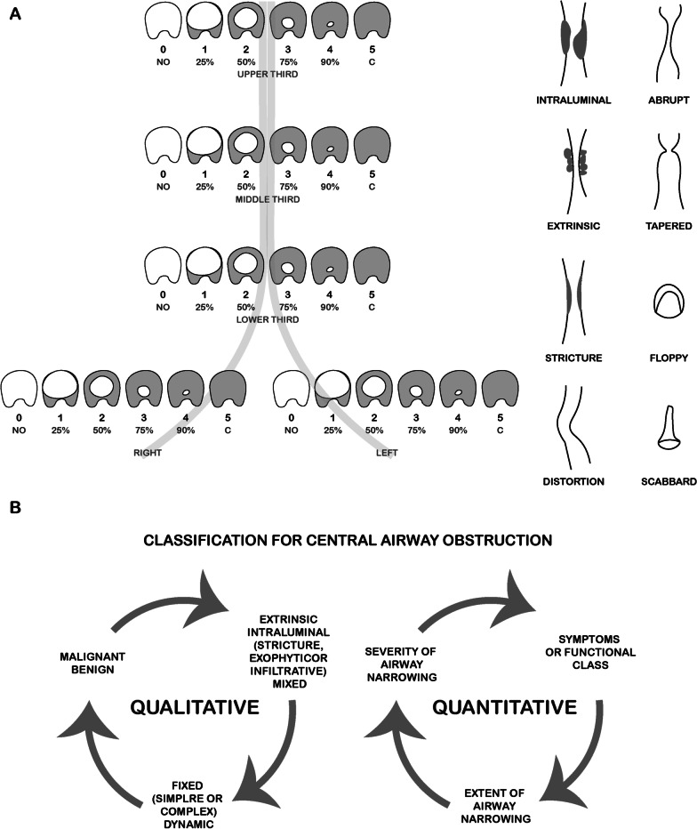
Schematic – Types of Central Airway Obstruction (Freitag/Murgu)

Specimen – Biodegradable stent fibers (coughed fragments)

Stenting – Metallic Y stent for bronchopleural fistula (LLL)

Stenting – Metallic Y stent for bronchopleural fistula (RML deployment sequence)

Stenting – Metallic Y stent for bronchopleural fistula (RML migration and successful reimplantation)

Stenting – Metallic Y stent removal (hook technique with radiography/CT confirmation)
Stenting –Distal Granulation at Endobronchial Stent

Stenting –Dumon Arm with Cut‑Out + Micro‑Tech “Stent‑in‑Stent”

Stenting –GINA Silicone Stent (Design/Anti‑Migration)

Stenting –GINA vs Dumon (Bench Tests: Anti‑Migration/Force/Flex)

Stenting –GINA vs Dumon (Porcine Stenosis Models; 21 Days)

Stenting –Hook‑Sheath Technique (Tracheal Stent Removal)
Stenting –Micro‑Tech Straight FC‑SEMS
Stenting –Neo‑epithelialization (Metal Stents; 4 Patients)

Stenting –Polydioxanone Tracheal Stent (Radiopaque Markers)

Stenting –Post‑Placement Confirmation (Bronchoscopy)

Stenting –SEMS Deploying System (Aero; Merit Endotek)

Stenting –Silicone Stent Deploying System (Polyflex)
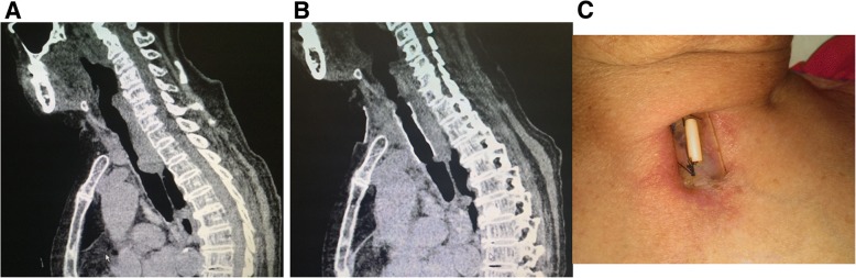
Stenting –Suture Fixation (Case #1; Sagittal CT & Neck View)
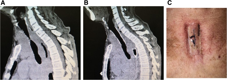
Stenting –Suture Fixation (Case #9; Sagittal CT & Neck View)
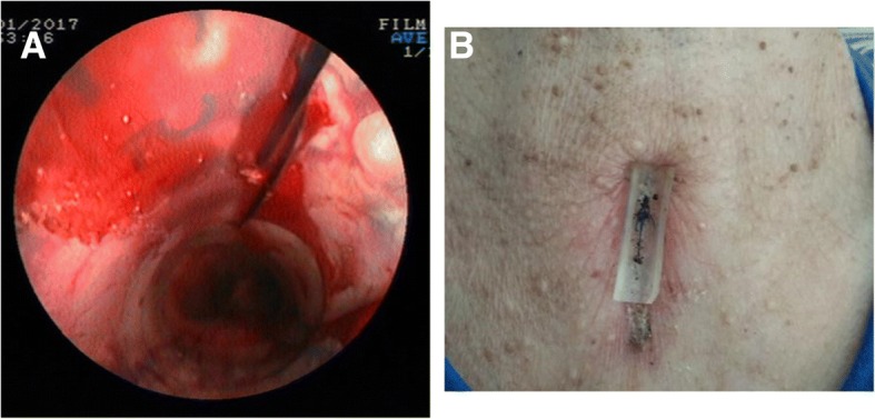
Stenting –Suture Fixation (Percutaneous Pad; Post‑Op CT)

Stenting –Through‑the‑Scope (TTS) vs Over‑the‑Wire (OTW) Design

Stenting –Y Silicone in Stomach (Migration Case)

Combined Airway/Esophageal Stents (TEF; Fluoro‑Guided)

Esophageal Stent Migration (Relocation Under Fluoro)

MWA Inside Silicone Stent (Ball‑Valve Tumor)
PDT (Early Lung Cancer; Fiber Alongside Lesion)

Post‑PDT Debris (Bronchoscopic View)

TEF Coverage with Self‑Expanding Y SEMS (CT/Bronch)

TPO (Carina/Main Bronchi)

TPO (Tracheobronchopathia Osteochondroplastica; Trachea)

Y‑SEMS for Severe Bilateral MAO (Recovery)

Stent – Through‑the‑scope (TTS) outcomes

Bronchoscopic Images – Laser Photocoagulation (YAP/Nd:YAG)
Fluoroscopy – Bilateral SEMS placement (side-by-side method, step-by-step)
Instrumentation – CoreCath 2.7S (Multimodal Debulking)

Direct Laryngoscopy – Subglottic stenosis (0-degree endoscope view)

CT/Spirometry – CAO pre/post stent (flow-volume loops, chemoradiotherapy response)

Airway Stenosis – Montgomery T-tube placement procedure

Algorithm/Procedure – iSGS treatment approaches (endoscopic laryngotracheoplasty techniques)